What Is The Muscular System? Anatomy And Physiology
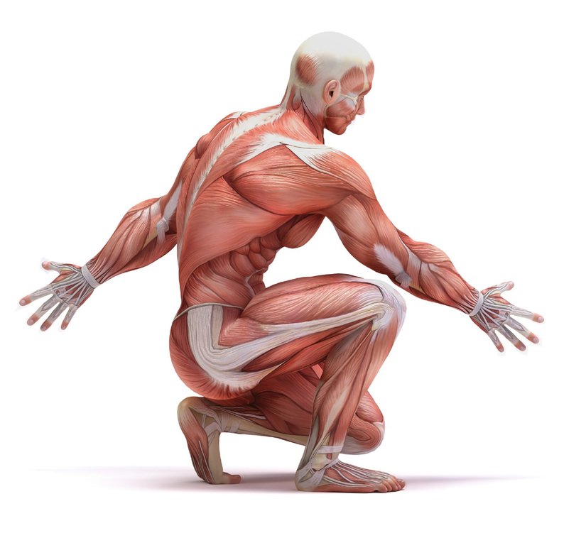
The muscular system is a complex network of tissues and organs that work together to facilitate movement, stability, and posture in the human body. Comprised of three types of muscles – skeletal, smooth, and cardiac – the muscular system plays a vital role in maintaining overall health and wellness.
Understanding the anatomy and physiology of the muscular system is critical for healthcare professionals, athletes, and anyone seeking to improve their physical fitness and wellbeing. In this article, we will explore the structure and function of each muscle type, as well as the physiology of muscle contraction and the role of calcium in muscle function.
Additionally, we will discuss common injuries and disorders of the muscular system, along with the importance of the neuromuscular junction in facilitating muscle movement.
By the end of this article, readers will have a comprehensive understanding of what the muscular system is, how it works, and why it is so important for overall health and wellbeing.
Key Takeaways
- The muscular system facilitates movement, stability, and posture in the human body and is composed of three types of muscles: skeletal, smooth, and cardiac.
- The neuromuscular junction is crucial for proper function of all muscles and is the site of communication between motor neuron and muscle fiber.
- Common injuries and disorders of the muscular system include muscle strains, sprains, fibromyalgia, muscular dystrophy, and myasthenia gravis.
- Understanding the muscular system is important for healthcare professionals, athletes, and anyone seeking to improve their physical fitness and wellbeing. Proper care and understanding of the muscular system is important for overall health and wellbeing.
The Three Types of Muscles in the Human Body
The human body is composed of three distinct types of muscles, including skeletal, smooth, and cardiac muscles.
Skeletal muscles are attached to bones and are responsible for voluntary movements such as walking, running, and lifting weights. They are also involved in maintaining posture and body position. Skeletal muscles are composed of long, cylindrical cells called muscle fibers, which contain many nuclei and are striated, giving them a striped appearance under a microscope.
Smooth muscles, on the other hand, are found in the walls of internal organs such as the stomach, intestines, and blood vessels. They are responsible for involuntary movements such as peristalsis, the wave-like contractions that move food through the digestive tract. Smooth muscles are non-striated and have a spindle-shaped appearance. They are controlled by the autonomic nervous system, which means that they function without conscious control.
The third type of muscle, cardiac muscle, is found only in the heart. It is also striated, like skeletal muscle, but it is involuntary, like smooth muscle. The unique properties of cardiac muscle allow it to contract rhythmically and efficiently pump blood throughout the body.
The Anatomy of Skeletal Muscles
Skeletal muscles are composed of muscle fibers that are bundled together to form fascicles. These fascicles are then surrounded by connective tissue, which is called the epimysium.
Within the fascicle, there are individual muscle fibers, which are surrounded by the perimysium. Finally, each muscle fiber is composed of myofibrils, which are made up of sarcomeres.
The sarcomere is the basic unit of muscle contraction and is made up of actin and myosin filaments. When a muscle contracts, the myosin filaments pull the actin filaments closer together, shortening the sarcomere and causing the muscle to contract.
This contraction is initiated by a signal from the nervous system, which travels down the motor neuron and releases a neurotransmitter called acetylcholine at the neuromuscular junction. This triggers a series of events that ultimately leads to the contraction of the muscle fiber.
Understanding the anatomy of skeletal muscles is crucial to understanding how they function and how they can be trained to improve performance.
The Physiology of Muscle Contraction
Actin and myosin filaments are the two main proteins involved in muscle contraction. These filaments are arranged in a specific pattern within the muscle fiber, with actin filaments forming a thin layer around the myosin filaments.
When a muscle fiber receives a signal from a motor neuron, calcium ions are released into the muscle cell, which bind to a protein called troponin. This binding causes a shift in the position of tropomyosin, which exposes the binding sites on the actin filaments. Myosin heads then attach to these binding sites, forming cross-bridges between the actin and myosin filaments.
Once the cross-bridges are formed, the myosin heads undergo a conformational change, pulling the actin filaments towards the center of the sarcomere. This movement is powered by the hydrolysis of ATP, which provides the energy needed for the myosin heads to detach from the actin filaments and reset for the next cycle of contraction.
The repeated formation and detachment of cross-bridges results in the shortening of the sarcomere, which in turn causes the muscle fiber to contract.
Understanding the complex processes involved in muscle contraction is essential for developing effective treatments for muscle disorders and injuries.
Muscle contraction is a complex process that involves the interaction of actin and myosin filaments, as well as the release of calcium ions and the hydrolysis of ATP.
The formation and detachment of cross-bridges between these filaments results in the shortening of the sarcomere and the contraction of the muscle fiber.
A thorough understanding of the physiology of muscle contraction is crucial for developing treatments for muscle disorders and injuries, as well as for improving athletic performance.
The Role of Calcium in Muscle Function
Calcium ions play a critical role in muscle function. When a motor neuron sends an electrical signal to a muscle fiber, calcium ions are released from the sarcoplasmic reticulum, a specialized type of endoplasmic reticulum found in muscle cells.
These calcium ions then bind to troponin, a protein found on the surface of actin filaments within muscle fibers. This binding causes a conformational change in tropomyosin, another protein located on the actin filament. The conformational change exposes the binding sites on actin, allowing myosin heads to attach and initiate muscle contraction.
In addition to triggering muscle contraction, calcium ions also play a role in muscle relaxation. After the electrical signal from the motor neuron ceases, calcium ions are actively pumped back into the sarcoplasmic reticulum by a calcium-ATPase pump.
As calcium ions are removed from troponin, tropomyosin blocks the binding sites on actin, preventing myosin heads from attaching and inhibiting muscle contraction. Therefore, the proper regulation of calcium ions is essential for the proper function of the muscular system.
The Structure and Function of Smooth Muscle
Smooth muscle is a type of muscle tissue that is composed of spindle-shaped cells with a single nucleus and lacks the striations seen in skeletal muscle. These muscles are found in the walls of various organs such as the digestive tract, blood vessels, and uterus. The smooth muscle cells are connected by gap junctions, which allow for coordinated contraction of the muscle fibers.
The structure and function of smooth muscle are different from skeletal muscle. Here are some key differences:
- Smooth muscle is controlled involuntarily, whereas skeletal muscle is controlled voluntarily. The contraction of smooth muscle is regulated by the autonomic nervous system and hormones.
- Smooth muscle can sustain contractions for longer periods of time than skeletal muscle. This is due to the presence of latch bridges, which keep the myosin and actin filaments bound together even when ATP is not available.
- Smooth muscle has a greater ability to stretch and maintain tension than skeletal muscle. This allows for the proper functioning of organs such as the bladder and uterus, which need to stretch and contract during certain physiological processes.
Overall, smooth muscle plays a crucial role in the proper functioning of many organs in the body. Understanding its unique structure and function is important in the treatment of various medical conditions that affect smooth muscle, such as asthma, hypertension, and uterine disorders.
The Unique Properties of Cardiac Muscle
Cardiac muscle, found exclusively in the heart, possesses unique properties that allow it to contract rhythmically and continuously to maintain circulation throughout the body. Unlike skeletal and smooth muscle, cardiac muscle cells are interconnected by intercalated discs, which contain gap junctions that allow for rapid electrical conduction and coordination of contraction. The cells also contain a large number of mitochondria, which provide the necessary energy for continuous contraction.
In addition, the cells of cardiac muscle have a longer refractory period than other types of muscle, which prevents tetanus and allows for relaxation and refilling of the heart chambers with blood. These unique properties of cardiac muscle are essential for maintaining proper circulation and preventing heart failure. The emotional impact of this fact can be represented in the following table:
| Fact | Emotional Response | ||
|---|---|---|---|
| Cardiac muscle allows for continuous circulation throughout the body | A sense of security and appreciation for the heart’s ability to keep us alive | ||
| The unique properties of cardiac muscle prevent heart failure | Gratitude for the intricacies of the body and the importance of taking care of our cardiovascular health | Recognizing the impact of lifestyle choices on heart health and making conscious efforts to maintain a healthy heart. |
The Importance of the Neuromuscular Junction
The previous subtopic discussed the unique properties of cardiac muscle. While it is true that the cardiac muscle possesses unique features that set it apart from the other types of muscle in the body, it is important to note that all muscles, regardless of their type, rely on the neuromuscular junction for proper function.
The neuromuscular junction is a specialized synapse between a motor neuron and a muscle fiber. It is the site at which the neuron communicates with the muscle fiber, allowing for the initiation of muscle contraction.
The importance of the neuromuscular junction lies in its ability to transmit information from the nervous system to the muscles. When a motor neuron is stimulated, it releases a neurotransmitter called acetylcholine, which binds to receptors on the muscle fiber and triggers a series of events that ultimately lead to muscle contraction.
Without this communication between the nervous system and the muscles, it would be impossible for our bodies to move and perform the countless tasks that we do every day. Therefore, understanding the anatomy and physiology of the neuromuscular junction is crucial for anyone interested in studying the muscular system.
Common Injuries and Disorders of the Muscular System
Injuries and disorders affecting the proper function of muscles can lead to significant impairments in daily activities and overall quality of life. One of the most common muscular injuries is a strain, which occurs when muscles are stretched beyond their capacity, causing damage to the muscle fibers. Strains can result from overuse, sudden movements, or inadequate warm-up before exercise. Symptoms of a strain include pain, swelling, and limited range of motion. Treatment typically involves rest, ice, compression, and elevation, followed by physical therapy to strengthen the affected muscle.
Another common muscular injury is a sprain, which occurs when ligaments, the bands of tissue that connect bones to each other, are stretched or torn. Sprains can happen when a joint is twisted or turned beyond its normal range of motion. Symptoms of a sprain include pain, swelling, bruising, and difficulty moving the affected joint. Treatment typically involves rest, ice, compression, and elevation, followed by physical therapy to help restore mobility and strength. In more severe cases, surgery may be necessary to repair the damaged ligament.
| Disorder/Injury | Description | Treatment | ||
|---|---|---|---|---|
| Fibromyalgia | A chronic disorder characterized by widespread musculoskeletal pain, fatigue, and tenderness in localized areas. | Medications, physical therapy, stress management techniques. | ||
| Muscular Dystrophy | A group of genetic disorders that cause progressive weakness and loss of muscle mass. | Physical therapy, assistive devices, medication for symptom relief. | ||
| Myasthenia Gravis | A chronic autoimmune disorder that affects the neuromuscular junction, causing muscle weakness and fatigue. | Medications, plasmapheresis, thymectomy (removal of the thymus gland). | Treatment aims to improve muscle strength and function, and may involve a combination of medication, physical therapy, and lifestyle modifications. |
Conclusion
In conclusion, the muscular system plays a vital role in maintaining bodily functions and movement. Comprised of three types of muscles, skeletal, smooth, and cardiac, each has unique properties and functions.
The anatomy and physiology of skeletal muscles involve the sliding filament theory and the neuromuscular junction, while smooth muscles are responsible for involuntary movements like digestion and cardiac muscles for the beating of the heart. However, the muscular system is not immune to injuries and disorders, such as strains, sprains, and muscular dystrophy, which can affect an individual’s quality of life. Understanding the anatomy and physiology of the muscular system can help in the prevention and treatment of these conditions.
Moreover, the muscular system also interacts with other systems in the body, such as the skeletal, nervous, and cardiovascular systems, to perform various functions. For example, the muscles work with bones to produce movement, receive signals from the nervous system to contract and relax, and receive oxygen and nutrients from the cardiovascular system.
Therefore, it is important to maintain a healthy muscular system through regular exercise, proper nutrition, and seeking medical attention when necessary. Overall, the muscular system is a complex and essential system that contributes to the overall health and well-being of an individual.

